Difference between revisions of "Moss Menageries with AGiR!"
| Line 17: | Line 17: | ||
Thanks to a number of inquiries, I finally found someone who could identify this being, which is actually a sort of amoeba, one of a number termed 'testate amoeba' which make a special protective 'helmet' of mineral matter (especially silica) and its own secreted 'glue.' | Thanks to a number of inquiries, I finally found someone who could identify this being, which is actually a sort of amoeba, one of a number termed 'testate amoeba' which make a special protective 'helmet' of mineral matter (especially silica) and its own secreted 'glue.' | ||
| − | George Sartiano is his name, a retired professor and medical doctor in oncology | + | George Sartiano is his name, a retired professor and medical doctor in oncology, and he was so inspired by my request, that he has made [https://www.youtube.com/channel/UCaaxbqgO8TepGzJbj99tV3g a new youtube channel for his video micrographs] (now with over 50 - check them out!)! |
| − | This is some of the info he passed on: | + | This is some of the info he passed on regarding the animal above (which had a population explosion in at least one of the moss menageries): |
| − | Order Arcellinida – a form of testate (shelled) amoeba.Testate amoebae have a “test” or tectum of their own making. The amoeba lives within the test, which is typically shaped like an inverted vase, with a single, terminal ventral aperture, through which the organism protrudes its pseudopodia (in your photomicrographs, the pseudopodia appear to be lobose in type). The test is often composed of mineral bits, sand granules, fragments and frustules of diatoms, etc., held together by a cement-like secretion of the amoeba ---sometimes giving the shell a tossed-together, hodge-podge like appearance. Various genera/species are more or less selective of the content of their shells, providing some basis for identification and classification. The nucleus of the organism is often visible through the shell; it usually takes on a spheroidal or round appearance; it may well be the dark, spherical object located centrally in your middle photomicrograph. Freshwater species often contain cytoplasmic organic symbionts – hence your observation of possible algal content is probably correct (photomicrographs #’s 1 and 3 illustrate this best).. | + | |
| − | Beyond identifying the organism’s Order, there are many genera and numerous species of Arcellinida. My initial reaction to your photomicrographs was to think that it is a difflugid (i.e., a species of difflugia). Difflugia are quite common in moist, watery habitats, and the descriptive literature does specifically mention mosses (re: your observation). | + | This protozoan is most likely of the 'Order Arcellinida – a form of testate (shelled) amoeba.Testate amoebae have a “test” or tectum of their own making. The amoeba lives within the test, which is typically shaped like an inverted vase, with a single, terminal ventral aperture, through which the organism protrudes its pseudopodia (in your photomicrographs, the pseudopodia appear to be lobose in type). The test is often composed of mineral bits, sand granules, fragments and frustules of diatoms, etc., held together by a cement-like secretion of the amoeba ---sometimes giving the shell a tossed-together, hodge-podge like appearance. Various genera/species are more or less selective of the content of their shells, providing some basis for identification and classification. The nucleus of the organism is often visible through the shell; it usually takes on a spheroidal or round appearance; it may well be the dark, spherical object located centrally in your middle photomicrograph. Freshwater species often contain cytoplasmic organic symbionts – hence your observation of possible algal content is probably correct (photomicrographs #’s 1 and 3 illustrate this best).. |
| + | Beyond identifying the organism’s Order, there are many genera and numerous species of Arcellinida. My initial reaction to your photomicrographs was to think that it is a difflugid (i.e., a species of difflugia). Difflugia are quite common in moist, watery habitats, and the descriptive literature does specifically mention mosses (re: your observation).' | ||
Revision as of 16:43, 3 November 2016
This will be the space to add info about the Moss Fauna that are being studied at the Hackuarium!! Glass petrie dishes with moss are available for all to examine! (below the bench, above the drawers, opposite the microscopes)
Some very interesting creatures, including rotifers, amoeba, and waterbears (tardigrades) have been spotted. The latest mini-menagerie added cultured waterbears from Vanessa to moss from Glion.
The idea behind this effort is to raise awareness of how many different species can be found in this natural habitat! Hopefully, this will aid in terms of public consciousness about how we impact these varied creatures without even realising it, and stimulate conservation efforts!
Amazingly, fully dry moss harbors many creatures that can be 'brought to life' when put in water! Following the evolution of these miniature ecosystems over time is also of great interest, so eventually this page will link to other pages following individual 'cultures' - some of which were established last summer!
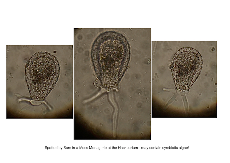
Just to begin, here is one special protozoan, first spotted on the move by Sam. (thanks!)
Thanks to a number of inquiries, I finally found someone who could identify this being, which is actually a sort of amoeba, one of a number termed 'testate amoeba' which make a special protective 'helmet' of mineral matter (especially silica) and its own secreted 'glue.'
George Sartiano is his name, a retired professor and medical doctor in oncology, and he was so inspired by my request, that he has made a new youtube channel for his video micrographs (now with over 50 - check them out!)!
This is some of the info he passed on regarding the animal above (which had a population explosion in at least one of the moss menageries):
This protozoan is most likely of the 'Order Arcellinida – a form of testate (shelled) amoeba.Testate amoebae have a “test” or tectum of their own making. The amoeba lives within the test, which is typically shaped like an inverted vase, with a single, terminal ventral aperture, through which the organism protrudes its pseudopodia (in your photomicrographs, the pseudopodia appear to be lobose in type). The test is often composed of mineral bits, sand granules, fragments and frustules of diatoms, etc., held together by a cement-like secretion of the amoeba ---sometimes giving the shell a tossed-together, hodge-podge like appearance. Various genera/species are more or less selective of the content of their shells, providing some basis for identification and classification. The nucleus of the organism is often visible through the shell; it usually takes on a spheroidal or round appearance; it may well be the dark, spherical object located centrally in your middle photomicrograph. Freshwater species often contain cytoplasmic organic symbionts – hence your observation of possible algal content is probably correct (photomicrographs #’s 1 and 3 illustrate this best).. Beyond identifying the organism’s Order, there are many genera and numerous species of Arcellinida. My initial reaction to your photomicrographs was to think that it is a difflugid (i.e., a species of difflugia). Difflugia are quite common in moist, watery habitats, and the descriptive literature does specifically mention mosses (re: your observation).'
