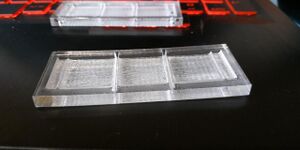
Chip for genomic integrity 2018
This chip prototype is part of the Genomic Integrity 2018.
Goals
As many steps of the comet cell assay should be carried out on the chip.
- Concentration of the cells by centrifugation
- Creation of the agarose pads
- Treatment of the agarose pads
- Electrophoresis
- Analyze of the agarose pads
The chip should be easy to produce (either considering the materials we use or the machinery). It should be reusable. As the chip's users won't be used to work in a lab, the chip will need to be easy to use and handle. The chip will be made for 3 pads in total. The original idea is to make one treated pad, one normal and one untreated, but the user will be able to change the conditions however he wants. We will make various slides, which will need to be changed according to the step.
Actual design(s)
Design 1 (improved)
We decided to make the chip the size of a microscope slide (25mm height, 75mm long), for it to be easily observable under a normal microscope.
In the end too thick to be observable under a microscope
Concentration of the cells by centrifugation
This step is not implemented in the device yet.
Creation of the agarose pads
For this first step, we thought about using 3 slides. A top one, a middle one and a bottom one.
- The top one contains 6 holes, 2 for each pad (entry and exit hole if overflow). This pads allows to make a really thin pad.
- The middle one is made out of Xerox transparencies paper (ref: 3R96002), and contain three round holes of 15mm diameter for the three pads (Squared pads tend to stick to the walls of the device afterwards)
- The bottom one is a normal slide
Treatment of the agarose pads & Electrophoresis
After having settled, the pads must be transferred into a larger well to allow them to float around in liquid. These new wells will be 20x20mm, larger than the size of a coverslip (otherwise the lasercutter makes them too small). For the electrophoresis, we need two electrodes. We also need the agarose pads to be in contact with the liquid.
- One bottom slide with 3 wells of 20x20mm (approximately 3.5mm deep), flanked by small walls (approximately 1.2mm deep, 1x20mm), and on the sides two smaller wells for the electrodes (3.5mm deep, 3x20mm)
Protocol : Put a coverslip in each of the 20x20mm well. Add some liquid on them. From the previous step, remove the top slide, and return the two others slide on this step bottom slide. Remove carefully the slides and push gently the pads into their new wells. Then perform the wanted treatment by changing the liquid in the well with a pipette or Paster's pipette. Do the treatment and then return the whole chip onto a slide and add things through the holes on the sides (electrodes & liquid)
Problem: the coverslips do not fall on the slide they stick to the chip
Analyze of the agarose pads
Just remove the electrophoresis chip, we should end up with a slide with the pads and coverslips on top. Maybe should find a way to dry or change the coverslip before microscope use.
Design 2
We decided to make the chip larger than the size of a microscope slide (25mm height, 75mm long), as anyway it will be too big to be observable under a microscope.
Concentration of the cells by centrifugation
This step is not implemented in the device yet.
Creation of the agarose pads
For this first step, we thought about using 3 slides. A top one, a middle one and a bottom one.
- The top one contains 6 holes, 2 for each pad (entry and exit hole if overflow). This pads allows to make a really thin pad.
- The middle one is made out of Xerox transparencies paper (ref: 3R96002), and contain three round holes of 15mm diameter for the three pads (Squared pads tend to stick to the walls of the device afterwards)
- The bottom one is a normal slide
Treatment of the agarose pads & Electrophoresis
After having settled, the pads must be transferred into a larger well to allow them to float around in liquid. These new wells will be 20x20mm, larger than the size of a coverslip (otherwise the lasercutter makes them too small). For the electrophoresis, we need two electrodes. We also need the agarose pads to be in contact with the liquid.
- One bottom slide with 3 wells of 20x20mm (approximately 2.5mm deep), separated by small deeper wells for removable walls (approximately 3.5mm deep, 1.5x26.5mm), and on the sides two smaller wells for the electrodes (2.5mm deep, 3x20mm)
Protocol : Put a slide vertically to make the removable walls. Put a coverslip in each of the 20x20mm well. Add some liquid on them. From the previous step, remove the top slide, and return the two others slide on this step bottom slide. Remove carefully the slides and push gently the pads into their new wells. Then perform the wanted treatment by changing the liquid in the well with a pipette or Paster's pipette. Do the treatment, remove the liquid, remove the walls and add the electrophoresis buffer. Then run the electrophoresis.
Analyze of the agarose pads
Just remove the electrophoresis chip, we should end up with a slide with the pads and coverslips on top. Maybe should find a way to dry or change the coverslip before microscope use.
Previous Designs
Design 1
We decided to make the chip the size of a microscope slide (25mm height, 75mm long), for it to be easily observable under a normal microscope. We will make various slides, which will need to be changed according to the step. The chip will be made for 3 pads in total. The original idea is to make one treated pad, one normal and one untreated, but the user will be able to change the conditions however he wants.
Concentration of the cells by centrifugation
This step is not implemented in the device yet.
Creation of the agarose pads
For this first step, we thought about using 3 slides. A top one, a middle one and a bottom one.
- The top one will contain 6 holes, 2 for each pad (entry and exit hole if overflow)
- The middle one will be thin (if possible 50µm or 100µm thick), and contain three holes of 15x15mm for the three pads
- The bottom one will be a full normal slide
To check: is the top pad really necessary
Treatment of the agarose pads
After having settled, the pads must be transferred into a larger well to allow them to float around in liquid. These new wells will be 18x18mm, the size of a coverslip for microscope slide.
- One bottom slide with 3 wells of 18x18mm, 3 times deeper than the agarose pads.
From the previous step, remove the top slide, and return the two others slide on this step bottom slide. Remove carefully the slides and push gently the pads into their new wells. Then perform the wanted treatment by changing the liquid in the well with a pipette or Paster's pipette.
To check: is a top slide needed to perform the treatments
Electrophoresis
To make an electrophoresis, we need two electrodes. We also need the agarose pads to be in contact with the liquid. The bottom slide for this test is the most complex of our slides.
- One bottom slide with three wells of 18x18mm, XXX µm deep, inside a bigger well less deep XXX µm, flanked by two deeper wells for the electrodes
- One top slide with two holes for the liquid and two for the electrodes. The electrodes will be attached to this slide. The holes will all be above the electrode's wells
Same thing as previous step return the slide containing the pads into this step's bottom slide.
Analyze of the agarose pads
The 18x18mm wells are the size of a common coverslip. Thus, removing the top of the chip, removing most liquid and adding a coverslip over the three 18x18mm wells should permit the observation of the pads under the microscope.
Challenges & Problems
- The chip does not for the moment enable the concentration of the cells into a small volume. We do perform two centrifugations in the lab, we must see if just letting the cells settle down by gravity would be enough.
- The chip slide would be easily observable under a microscope, but maybe more challenging to do a slide observable with a DIY microscope (like a Foldscope).
- The chip requires movement of the pads from one slide to another, maybe we should at least label the slides a certain way to make sure not to mix the pads by inadvertence.

